
Page 93 - Demo
P. 93
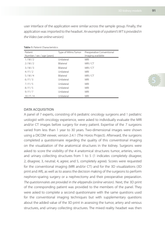
3Dkidneymodels915userinterfaceoftheapplicationweresimilaracrossthesamplegroupFinallytheapplication wasimported to theheadset Anexampleofa patient’sWTis providedintheVideo(seeonlineversion)Table1PatientCharacteristicsPatientNumber /sex /age(years)Typeof Wilms TumorPreoperativeConventionalImagingavailable1 /M /2UnilateralMRI2 /M /2BilateralMRI /CT3 /M /3BilateralMRI /CT4 /F /2UnilateralMRI5 /M /4BilateralMRI /CT6 /F /3UnilateralMRI7 /F /5UnilateralMRI8 /F /5UnilateralMRI9 /F /7UnilateralMRI10 /F /4UnilateralMRIDATAACQUISITIONApanelof7expertsconsistingof6pediatriconcologysurgeonsand1pediatricurologistwithoncologyexperiencewereaskedtoindividuallyevaluatetheMRIand/orCTimagesbeforesurgeryforeverypatientExpertiseofthe7surgeonsvariedfromlessthan1yearto30yearsTwo-dimensionalimageswereshownusingaDICOMviewer,version241(TheHorosProject)AfterwardthesurgeonscompletedaquestionnaireregardingthequalityofthisconventionalimagingonthevisualizationoftheanatomicalstructuresinthekidneySurgeonswereaskedtoscorethevisibilityofthe4anatomicalstructurestumor,arteriesveinsandurinarycollectingstructuresfrom1to5(1indicatescompletelydisagree;2disagree;3neutral;4agree;and5completelyagree)Scoreswererequestedfortheconventionalimaging(MRIand/orCT)andforthe3Dvisualizations(3DprintandAR)aswellastoassessthedecisionmakingofthesurgeonstoperformnephron-sparingsurgeryoranephrectomyandtheirpreoperativepreparationThequestionnairesareprovidedintheeAppendix(onlineversion)Nextthe3DprintofthecorrespondingpatientwasprovidedtothemembersofthepanelTheywereaskedtocompleteasecondquestionnairewiththesamequestionsusedfortheconventionalimagingtechniquesbutwithsupplementaryquestionsabouttheaddedvalueofthe3Dprintinassessingthetumor,arteryandvenousstructuresandurinarycollectingstructuresThemixedrealityheadsetwasthen
