
Page 92 - Demo
P. 92
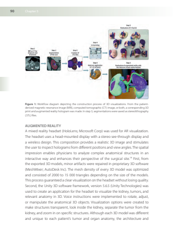
90Chapter5Figure1Workflowdiagramdepictingtheconstructionprocessof3DvisualizationsFromthepatientderivedmagneticresonanceimage(MRI)computedtomographic(CT)imageorbothacorresponding3DprintandaugmentedrealityhologramwasmadeInstep3segmentationsweresavedasstereolithography(STL)filesAUGMENTEDREALITYAmixedrealityheadset(HoloLens;MicrosoftCorp)wasusedforARvisualizationTheheadsetusesahead-mounteddisplaywithastereosee-throughdisplayandawirelessdesignThiscompositionprovidesarealistic3Dimageandstimulatestheuser toinspectholograms fromdifferentpositionsand viewangles Thespatialimpressionenablesphysicianstoanalyzecomplexanatomicalstructuresinaninteractivewayandenhancestheirperspectiveofthesurgicalsite18Firstfromtheexported3Dmodelsminorartifactswererepairedinproprietary3Dsoftware(MeshMixer;AutoDeskInc)Themeshdensityofevery3Dmodelwasoptimizedandconsistedof2000to15000trianglesdependingonthesizeofthemodelsThisprocess guaranteed a clear visualization on theheadset withoutlosing qualitySecondtheUnity3Dsoftwareframeworkversion565(Unity Technologies)wasusedtocreateanapplicationfortheheadsettovisualizethekidneytumorsandrelevantanatomyin3DVoiceinstructionswereimplementedtorotateadjustormanipulatetheanatomical3DobjectsVisualizationoptionswerecreatedtomakestructurestransparentlookinsidethekidneyseparatethetumorfromthekidneyandzoominonspecificstructuresAlthougheach3Dmodelwasdifferentanduniquetoeachpatient’stumorandorgananatomythearchitectureand
