
Page 36 - Demo
P. 36
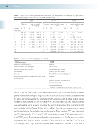
34Chapter2Table1Male(M)andFemale(F)distributionwithmeanageandstandarddeviationperagegroupfor3Dphotographs(3DP)CThardtissue(CT-HT)andCTsofttissue(CT-ST)3DPCT-HTCT-STNumberAge(months)mean(SD)NumberAge(months)mean(SD)nAge(months)mean(SD)Group(months)MFMFMFMFMFMF3141231(04)30(02)3723(12)20(16)3823(12)20(13)6101264(07)59(03)2155(07)60()1060()9131490(04)90(02)5194(09)90()3197(06)90()12109120(04)120(02)36117(12)125(05)36117(12)122(08)1547152(03)151(05)55154(09)148(08)44153(10)153(05)18108185(08)182(01)25195(21)192(15)44198(15)193(17)2434243(03)242(01)76271(28)248(22)75266(25)244(23)361712362(30)360(28)108371(26)365(31)4852456(48)445(07)32477(55)445(07)Table2Landmarkson3Dphotographsand3D-CT.3Dphotographs3DCTPretragion(leftandright)NasionLateralcanthus(leftandright)SellaturcicaMedialcanthus(leftandright)FrontozygomaticsutureNasalbridgeExternalacousticmeatusNosetipFrontalintersectionofthepterionSubnasallandmarkatthetransitionofthenoseandupperlipAsterionExternaloccipitalprotuberanceAnteriorfontanellePosteriorfontanelleAdditionallandmarksovercoronalsuturesn=8ofthecranium Thesetwopointswereusedtoindicateahorizontalmeasurementplane on the cranial shapes(Figure 2) Themaximum cranial width was determinedbyalineperpendiculartothecephaliclengthCranialwidthdividedbythecraniallengthandmultipliedby100resultedinthecranialindex(CI) Thecircumferencewascalculatedusingaplanecrossingthepointsthatdefinethecephaliclengthandcephalicwidth(Figure2)Forallmodalitiesthevolumemeasurementswereperformedabovethesellaturcica–nasionplane Totalvolume(TV)wasmeasuredon3DphotographsCT-HT,andCT-ST.Intracranialvolume(ICV)wasmeasuredinthe CT-HT group The anterior andposterior components of the TV were computedseparatelyanddividedatthepositionofthesellaturcicaOntheCT-HTscansthemetopicandsagittalsuturelengthsweremeasuredoverthesurfaceinthe
