
Page 34 - Demo
P. 34
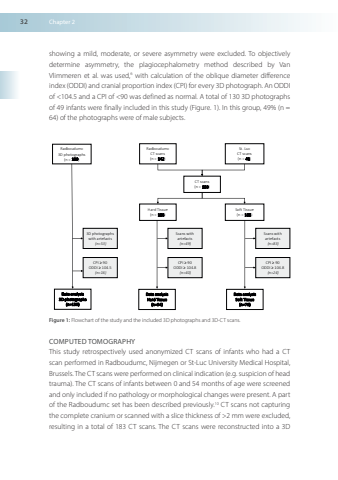
32Chapter2showingamildmoderateorsevereasymmetrywereexcludedToobjectivelydetermineasymmetrytheplagiocephalometrymethoddescribedbyVanVlimmerenetalwasused6withcalculationoftheobliquediameterdifferenceindex(ODDI)andcranialproportionindex(CPI) forevery3DphotographAnODDIof<1045andaCPIof<90wasdefinedasnormalAtotalof1303Dphotographsof49infantswerefinallyincludedinthisstudy(Figure1)Inthisgroup49%(n=64)ofthephotographswereofmalesubjectsRadboudumc3Dphotographs(n=199)RadboudumcCTscans(n=142)Scanswithartefacts(n=49)StLucCTscans(n=41)CPI≥90ODDI≥1048(n=40)HardTissue(n=183)SoftTissue(n=183)DataanalysisHardTissue(n=94)Dataanalysis3Dphotographs(n=130)3Dphotographswithartefacts(n=53)CPI≥90ODDI≥1045(n=16)Scanswithartefacts(n=83)CPI≥90ODDI≥1048(n=24)DataanalysisSoftTissue(n=76)CTscans(n=183)Figure1Flowchartofthestudyandtheincluded3Dphotographsand3D-CTscansCOMPUTED TOMOGRAPHYThisstudyretrospectivelyusedanonymizedCTscansofinfantswhohadaCTscanperformedinRadboudumcNijmegenorSt-LucUniversityMedicalHospitalBrussels The CTscans wereperformedonclinicalindication(egsuspicionofheadtrauma) TheCTscansofinfantsbetween0and54monthsofagewerescreenedandonlyincludedifnopathologyormorphologicalchangeswerepresentApartoftheRadboudumcsethasbeendescribedpreviously10CTscansnotcapturingthecompletecraniumorscannedwithaslicethicknessof>2mmwereexcludedresultinginatotalof183CTscansTheCTscanswerereconstructedintoa3D
