
Page 96 - Development of Functional Scaffolds for Bone Tissue Engineering Using 3D-Bioprinting of Cells and Biomaterials - Yasaman Zamani
P. 96
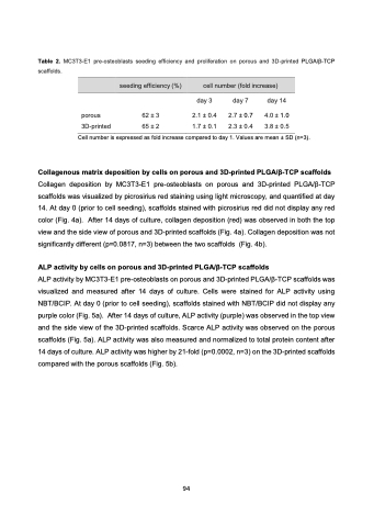
Table 2. MC3T3-E1 pre-osteoblasts seeding efficiency and proliferation on porous and 3D-printed PLGA/β-TCP scaffolds.
         seeding efficiency (%) cell number (fold increase)
      day 3
porous 62 ± 3 2.1 ± 0.4
3D-printed 65 ± 2 1.7 ± 0.1
day 7
2.7 ± 0.7
2.3 ± 0.4
day 14
4.0 ± 1.0
3.8 ± 0.5
Cell number is expressed as fold increase compared to day 1. Values are mean ± SD (n=3).
Collagenous matrix deposition by cells on porous and 3D-printed PLGA/β-TCP scaffolds
Collagen deposition by MC3T3-E1 pre-osteoblasts on porous and 3D-printed PLGA/β-TCP scaffolds was visualized by picrosirius red staining using light microscopy, and quantified at day 14. At day 0 (prior to cell seeding), scaffolds stained with picrosirius red did not display any red color (Fig. 4a). After 14 days of culture, collagen deposition (red) was observed in both the top view and the side view of porous and 3D-printed scaffolds (Fig. 4a). Collagen deposition was not significantly different (p=0.0817, n=3) between the two scaffolds (Fig. 4b).
ALP activity by cells on porous and 3D-printed PLGA/β-TCP scaffolds
ALP activity by MC3T3-E1 pre-osteoblasts on porous and 3D-printed PLGA/β-TCP scaffolds was visualized and measured after 14 days of culture. Cells were stained for ALP activity using NBT/BCIP. At day 0 (prior to cell seeding), scaffolds stained with NBT/BCIP did not display any purple color (Fig. 5a). After 14 days of culture, ALP activity (purple) was observed in the top view and the side view of the 3D-printed scaffolds. Scarce ALP activity was observed on the porous scaffolds (Fig. 5a). ALP activity was also measured and normalized to total protein content after 14 days of culture. ALP activity was higher by 21-fold (p=0.0002, n=3) on the 3D-printed scaffolds compared with the porous scaffolds (Fig. 5b).
 94
