
Page 98 - Development of Functional Scaffolds for Bone Tissue Engineering Using 3D-Bioprinting of Cells and Biomaterials - Yasaman Zamani
P. 98
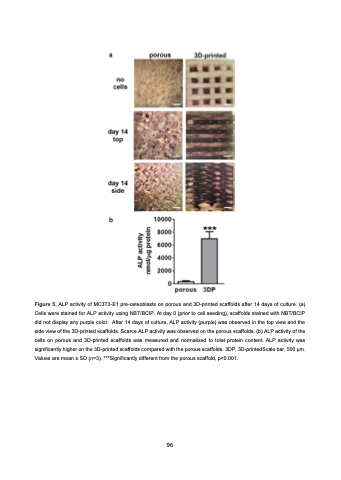
 Figure 5. ALP activity of MC3T3-E1 pre-osteoblasts on porous and 3D-printed scaffolds after 14 days of culture. (a) Cells were stained for ALP activity using NBT/BCIP. At day 0 (prior to cell seeding), scaffolds stained with NBT/BCIP did not display any purple color. After 14 days of culture, ALP activity (purple) was observed in the top view and the side view of the 3D-printed scaffolds. Scarce ALP activity was observed on the porous scaffolds. (b) ALP activity of the cells on porous and 3D-printed scaffolds was measured and normalized to total protein content. ALP activity was significantly higher on the 3D-printed scaffolds compared with the porous scaffolds. 3DP, 3D-printedScale bar, 500 μm. Values are mean ± SD (n=3). ***Significantly different from the porous scaffold, p<0.001.
96
