
Page 97 - Development of Functional Scaffolds for Bone Tissue Engineering Using 3D-Bioprinting of Cells and Biomaterials - Yasaman Zamani
P. 97
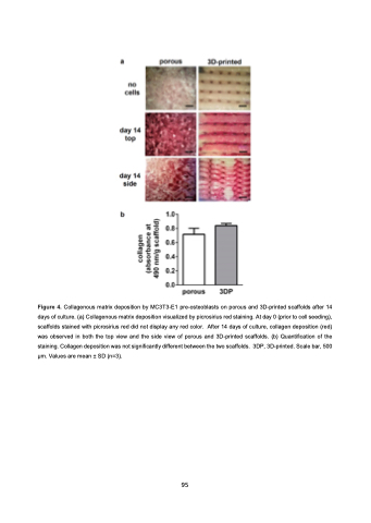
 Figure 4. Collagenous matrix deposition by MC3T3-E1 pre-osteoblasts on porous and 3D-printed scaffolds after 14 days of culture. (a) Collagenous matrix deposition visualized by picrosirius red staining. At day 0 (prior to cell seeding), scaffolds stained with picrosirius red did not display any red color. After 14 days of culture, collagen deposition (red) was observed in both the top view and the side view of porous and 3D-printed scaffolds. (b) Quantification of the staining. Collagen deposition was not significantly different between the two scaffolds. 3DP, 3D-printed. Scale bar, 500 μm. Values are mean ± SD (n=3).
95
