
Page 95 - Development of Functional Scaffolds for Bone Tissue Engineering Using 3D-Bioprinting of Cells and Biomaterials - Yasaman Zamani
P. 95
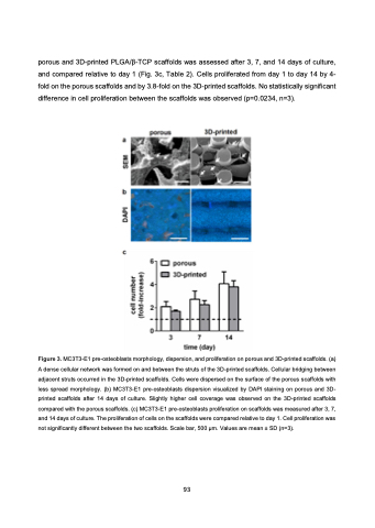
porous and 3D-printed PLGA/β-TCP scaffolds was assessed after 3, 7, and 14 days of culture, and compared relative to day 1 (Fig. 3c, Table 2). Cells proliferated from day 1 to day 14 by 4- fold on the porous scaffolds and by 3.8-fold on the 3D-printed scaffolds. No statistically significant difference in cell proliferation between the scaffolds was observed (p=0.0234, n=3).
Figure 3. MC3T3-E1 pre-osteoblasts morphology, dispersion, and proliferation on porous and 3D-printed scaffolds. (a) A dense cellular network was formed on and between the struts of the 3D-printed scaffolds. Cellular bridging between adjacent struts occurred in the 3D-printed scaffolds. Cells were dispersed on the surface of the porous scaffolds with less spread morphology. (b) MC3T3-E1 pre-osteoblasts dispersion visualized by DAPI staining on porous and 3D- printed scaffolds after 14 days of culture. Slightly higher cell coverage was observed on the 3D-printed scaffolds compared with the porous scaffolds. (c) MC3T3-E1 pre-osteoblasts proliferation on scaffolds was measured after 3, 7, and 14 days of culture. The proliferation of cells on the scaffolds were compared relative to day 1. Cell proliferation was not significantly different between the two scaffolds. Scale bar, 500 μm. Values are mean ± SD (n=3).
 93
