
Page 66 - Demo
P. 66
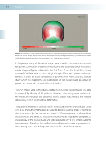
64Chapter3Figure 6 The fourPC’s which wereselected toidentify the trigonocephalypatients wereused tomanipulatethemeancranialshape TheresultedcranialshapedemonstratestemporalnarrowingandareducedorbitalwidthColoursindicateavolumeincrease(green)oravolumedecrease(red)InthepresentstudyallthecranialshapeswerescaledtothesamesizetocorrectforgrowthAlimitationofscalinginthisstudyistheassumptionthatthenormalcranialshapewillgrowuniformlyinthefirst3and6monthsInadditionitwasassumed that therewerenomorphologicalshapedifferencesbetweenmalesandfemalesInordertomakecomparisonofpatientsevenmoreaccurateafuturestudywhichinvestigatesthe3Dmodificationofthecranialshapeasaresultofgrowthandsexwouldbeavaluablecontribution8ThePCAmodelusedinthisstudycreatedfromnormalcranialshapeswasabletosuccessfullyidentifyall40patientsHowever,introducingmorevariationinthemodelbyincludingalsoabnormalcranialshapesmayimprovethemodel’srobustnesseveninseverecranialdeformitiesThepresentedmethodis a clinical tool for the evaluation of the cranial shapeUsingonlya3DphotothismethodcanbeusedtodetectifacranialshapeisnormalorabnormalinanobjectivemannerIncontrast to2Dmeasurementssuchascallipermeasurementsandother2Dmeasurementsthismodelregardsthecomplete3Dmorphologyofthecranialshapeandyetitproducesonlyafewsimpleoutcomemeasurements Therefore thismethodisanadditionandamajorimprovement tothecurrentlyusedclinicaldiagnosticmethodsforcranialabnormalities
