
Page 120 - Demo
P. 120
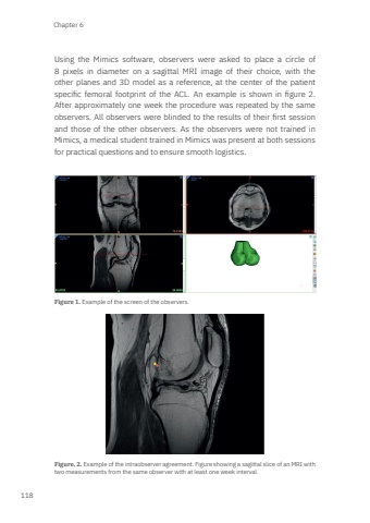
Chapter 6118Using the Mimics software, observers were asked to place a circle of 8 pixels in diameter on a sagittal MRI image of their choice, with the other planes and 3D model as a reference, at the center of the patient specific femoral footprint of the ACL. An example is shown in figure 2. After approximately one week the procedure was repeated by the same observers. All observers were blinded to the results of their first session and those of the other observers. As the observers were not trained in Mimics, a medical student trained in Mimics was present at both sessions for practical questions and to ensure smooth logistics.Figure 1. Example of the screen of the observers. Figure. 2. Example of the intraobserver agreement. Figure showing a sagittal slice of an MRI with two measurements from the same observer with at least one week interval. Mark Zee.indd 118 03-01-2024 08:56
