
Page 48 - Fluorescence-guided cancer surgery
P. 48
46
Chapter 3
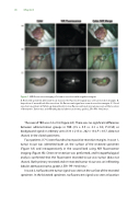
Figure 1. NIR uorescence imaging of a tumor resection with negative margins
A. Resected specimen after wide local excision. No uorescent signal was seen at resection margins. B. Inspection of wound bed after resection. No uorescent signal was seen at resection margins. C. Sliced resection specimen at Pathology department. A clear uorescent spot (arrow) was seen at the location of the tumor. Tumor was an in ltrating ductal adenocarcinoma, grade 2, ER+ PR+ Her2/neu-.
The overall TBR was 2.4 ± 0.8 (Figure 2A). There was no signi cant di erence between administration groups in TBR (2.5 ± 0.9 vs. 2.3 ± 0.5; P=0.50) or background signal in arbitrary units (333 ± 215 vs. 262 ± 134; P = 0.37, data not shown) in the sliced specimens.
Four patients (17%) were found to have positive resection margins. In case 1, tumor tissue was identi ed both on the surface of the resected specimen (Figure 3A) and intraoperatively in the wound bed using NIR uorescence imaging (Figure 3B). Direct re-resection was performed, and histopathological analysis con rmed that the uorescent resected tissue was tumor (data not shown). Both primary resected and re-resected tumor tissue was an in ltrating lobular adenocarcinoma, grade 2, ER+ PR- Her2/neu -.
In case 2, no uorescent tumor signal was seen on the surface of the resected specimen. In the bisected specimen, no uorescent signal was seen at location
