
Page 49 - Fluorescence-guided cancer surgery
P. 49
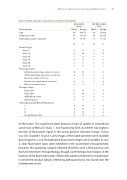
Table 2. Patient and tumor characteristics related to detectability
Detection of breast cancer using methylene blue 47
Detectable
N=20 N=4
Not Detectable
Characteristic
Age
Body mass index Pathological tumor size (mm)
Clinical Stage
Stage 0 Stage 1A Stage 1B Stage 2A Stage 2B Stage 3C
Histological type
In ltrating ductal type adenocarcinoma In ltrating lobular type adenocarcinoma Mucinous adenocarcinoma
Primary mucoepidermoid carcinoma Ductal carcinoma in situ
Receptor status
ER positive
PR positive HER2/NEU positive
Triple Negative
Histological grade (Bloom-Richardon)
I
II
III
No grading possible
Mean Range
58 (44-71) 26 (19-37) 16 (6-33)
Mean Range
68 (56-82) 24 (22-26) 10 (7-12)
N%N%
28 1 4 10 42 3 13 1 4 0 0 4 17 0 0 2 8 0 0 1 4 0 0
14 59 1 4 4 17 0 0 0 0 1 4 0 0 1 4 2 8 1 4
17 71 2 8 9 38 2 8 1 4 0 0 2 4 0 0
4 17 1 4 9 37 0 0 3 13 1 4 4 17 2 8
of the tumor. This could have been because of lack of uptake or intracellular conversion of MB to its leuco 11, non- uorescing form, but either way explains the lack of  uorescent signal in the tumor-positive resection margin. Tumor was a DCIS grade 3. In case 3, only images of the sliced specimen were available due to logistics, so no intraoperative  uorescent images were available. In case 4, clear  uorescent spots were identi ed in the wound bed intraoperatively, however, the operating surgeon believed that they were a false positive and did not resect them. Histopathology, though, con rmed positive margins at the location of the  uorescent spots. Afterwards, patient underwent a mastectomy in which the residual lobular in ltrating adenocarcinoma was found near the lumpectomy cavity.
