
Page 143 - Fluorescence-guided cancer surgery
P. 143
Imaging FRα positive ovarian and breast cancer 141
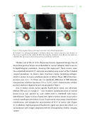
Figure 3. False-negative uorescent signal caused by a lack of depth penetration
A. Example of a metastasized greater omentum, which was clinical suspicious, but showed no uorescence signal from the outside. B. After dissecting the omentum, strong uorescent signal was identi ed. This observation shows the lack of tissue penetration at 500nm.
Thirteen out of the 57 (23%) uorescent lesions appeared benign. Five of these false positive lesions were identi ed as normal fallopian tube tissue on histopathological evaluation, showing FRα expression. These lesions were thus expected to bind EC17, and were resected anyway as part of the standard surgical procedure. Six lesions were structures mainly containing collagen, which is known to cause auto uorescence at 500nm. Mean TBR of the false- positives was 5.4 ± 1.0. There was no signi cant di erence in TBR between true-positives and false-positives (7.0 vs. 5.4; P = 0.47). Characteristics of false positive and false negative lesions are provided in Table 3.
Also in breast cancer, tumor-speci c uorescence signal was observed. Median TBR was 2.3 (range 2.1 – 6.2). However, auto uorescence of normal breast tissue was present to such extent that it interfered with tumor identi cation. Figure 4 shows uorescent signal in breast cancer tissue and in normal, auto uorescent breast tissue. Fluorescence microscopy showed clear membranous and cytoplasmic accumulation of EC17 in tumor cells (Figure 5). In addition, high background uorescent signal was observed, which is in concordance with images obtained with the intraoperative Artemis imaging system.
