
Page 141 - Fluorescence-guided cancer surgery
P. 141
Imaging FRα positive ovarian and breast cancer 139
Safety
All patients received 0.1 mg/kg EC17 over 10 minutes, and no infusion was intermitted or stopped. Infusion of EC17 was associated with mild, self-limiting hypersensitivity reactions in 7 out of 15 patients. The symptoms consisted of abdominal discomfort, itching throat and sneezing (for a summary list of treatment related adverse events, see Table S1, online available). One patient vomited after EC17 administration and received ondansetron 8 mg intravenously, followed by the planned surgical procedure. There were no clinical relevant changes in blood pressure or pulse rate compared to baseline.
Intraoperative uorescence imaging
Intraoperative uorescence imaging in ovarian cancer patients allowed clear detection of ovarian cancer lesions. Figure 1 shows an example of uorescent ovarian cancer metastases.
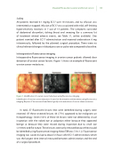
Figure 1. Identi cation of ovarian cancer metastases using uorescence imaging
Identi cation of ovarian cancer metastases located on the intestine and mesentery using uorescence
imaging. Biopsies of lesions were found histologically to be metastases of serous adenocarcinoma.
In total, 57 uorescent lesions that were identi ed during surgery were resected. Of these resected lesions 44 (77%) appeared to be malignant on histopathology. Seven (16%) of these 44 lesions were not detected by visual inspection with the naked eye or palpation either because they appeared benign or because they were missed during inspection due to small size (<10mm) and at nature. These lesions were only removed because these could be identi ed using uorescence imaging. Mean TBR was 7.0 ± 1.2. Fluorescence imaging was successful up to about 5.5 hours after EC17 administration, which was the longest time interval measured between administration and the end of a surgical procedure.
