
Page 145 - Fluorescence-guided cancer surgery
P. 145
Imaging FRα positive ovarian and breast cancer 143
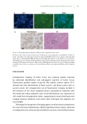
Figure 5. Histopathological evaluation of uorescence signal in breast cancer
A. Fluorescence microscopy showed clear membranous and cytoplasmic accumulation of EC17 in tumor cells. Blue color represents cell nuclei stained with DAPI, uorescence signal is indicated with green. Also a relatively high di usely uorescent background signal is seen, which is in concordance with the uorescence images obtained with the intraoperative imaging system. B. Immunohistological staining for FRα expression shows a FRα positive in ltrating breast cancer of no special type (example in dashed circle). Clear concordance is observed between uorescent signal and FRα positive malignant lesions.
DISCUSSION
Intraoperative imaging of tumor tissue may improve patient outcome by enhanced identi cation and subsequent resection of tumor tissue. Fluorescence guided surgery using the FRα speci c contrast agents EC17 allowed real time identi cation of both ovarian- and breast cancer cells. In ovarian cancer, the intraoperative use of uorescence imaging resulted in the resection of 16% more malignant lesions compared to inspection with the naked eye and/or palpation only. Visual identi cation was improved on stills made from intraoperative videos, supporting the notion that uorescent imaging improves detection even when other techniques like palpation are not available.
The biophysical properties of imaging agents are of paramount importance for successful tumor identi cation. Ideally, high uorescence signal is observed in malignant lesions, while normal or healthy tissue shows minimal uorescence
