
Page 142 - Fluorescence-guided cancer surgery
P. 142
140
Chapter 9
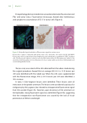
Histopathology demonstrated clear concordance between uorescence and FRα- and tumor status. Fluorescence microscopy showed clear membranous and cytoplasmic accumulation of EC17 in tumor cells (Figure 2).
Figure 2. Histopathological evaluation of uorescence signal in ovarian cancer
Fluorescence signal is indicated with green, blue color represents cell nuclei stained with DAPI. Fluorescence microscopy showed clear membranous and cytoplasmic accumulation of EC17 in tumor cells .The uorescent signal is located on all sites that stain positive for FRα expression, which is the anatomical site that appears to be a metastasis of serous ovarian adenocarcinoma on hematoxylin and eosin staining (dashed circle).
The (ex-vivo) assessment of the stills obtained from the videos made during the surgical procedure showed that on average (SD) 23.3 (± 11.9) lesions per still were identi ed with the naked eye. When the stills were supplemented with the uorescence image, 39.6 (± 22.7) lesions per still were identi ed, a 70% increase.
In total, 3 false-negative lesions were identi ed. These lesions were all metastases in the greater omentum. The lesions were considered suspicious for malignancy by the surgeons, but showed no intraoperative uorescence signal from the outside (Figure 3A). However, upon dissection of the omentum on the backtable, strong uorescent signal was identi ed (Figure 3B), suggesting that the intraoperative non- uorescence was caused by the lack of tissue penetration at 500nm wavelength.
