
Page 124 - Fluorescence-guided cancer surgery
P. 124
122
Chapter 8
All 8 histologically proven malignant metastatic lesions were NIR uorescent, so detection of metastatic lesions of ovarian cancer with ICG had a sensitivity of 100% in this study (Table 2). The speci city of NIR uorescence imaging could not be calculated, since lesions that were neither clinically suspect nor uorescent were not resected.
Table 2. Characteristics lesions found with ICG NIR uorescence imaging
NIR uorescent lesions
Concordance histopathology
True-positive False-positive
Ovarian / tubal carcinomas with metastatic disease N=2
8
N (%)
8 (100) 0 (0)
Ovarian / tubal carcinomas with non- metastatic disease N=4
9
N (%)
0 (0) 9 (100)
Endometrium carcinoma N=1
2
N (%)
0 (0) 2 (100)
Benign N=3
2
N (%)
0 (0) 2 (100)
Total N=10
21
N (%)
8 (38) 13 (62)
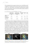
Clinically none of these 8 malignant and uorescent lesions had a benign appearance; therefore the use of NIR uorescence did not lead to the detection of otherwise undetected malignant lesions. In addition 13, on histological assessment, non-malignant lesions were also NIR uorescent, resulting in a false-positives rate of 62% (Table 2). Of these lesions, 2 were clinically characterized as malignant, 6 as suspicious for malignancy, and 5 as not suspicious for malignancy. Figure 4 shows an example of a NIR uorescent lesion that was found to be histologically benign, thus a false-positive lesion. This particular lesion was a calci ed lymph node. The localization, clinical appearance, pathology, and TBR for each false-positive lesion are listed in Table 3. Globally these false-positive lesions can be divided into two groups: normal tissue (N=10) and tissue with reactive changes (N=3).
Figure 4. Identi cation of ovarian cancer omental metastases using NIR uorescence imaging
Identi cation of a NIR uorescent lesion located in the mesentery of the intestine. The lesion was classi ed clinically as a metastasis but was found histologically to be a calci ed lymph node.
