
Page 123 - Fluorescence-guided cancer surgery
P. 123
Imaging based on the EPR e ect in ovarian cancer 121
show 2 NIR uorescent lesions located in the greater omentum of the same patient as presented in Figure 2, both containing serous adenocarcinoma.
Figure 2. Identi cation of ovarian cancer metastases using NIR uorescence imaging
A. Identi cation of ovarian cancer metastases located in a lymph node next to the right iliac vein (arrow) using NIR uorescence imaging. The lesion was found histologically to be a metastasis of serous adenocarcinoma. B. Ex vivo imaging of the same ovarian cancer metastases located in a lymph node next to the right iliac vein (arrow).
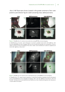
Figure 3. Identi cation of ovarian cancer omental metastases using NIR uorescence imaging
A. Identi cation of 2 ovarian cancer metastases located in the greater omentum (arrow and dashed arrow) using NIR uorescence imaging. B. Imaging of the same two NIR uorescent lesions removed from the omentum (arrow and dashed arrow). Both lesions were found histologically to be metastases of serous adenocarcinoma.
