
Page 122 - Fluorescence-guided cancer surgery
P. 122
120
Chapter 8
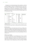
Histological assessment by the pathologist of the resected lesions con rmed the following diagnosis: 6 patients were diagnosed with either ovarian cancer (5) or cancer of the fallopian tube (1), of which the following subtypes were diagnosed: serous (3), clear-cell (1), endometrioid (1), mixed (1); one patient was diagnosed with endometrial cancer (endometriod type); and 3 patients had benign ovarian tumors. An overview of the nal histological diagnoses and FIGO stage is given in Table 1.
Table 1. Patient and tumor characteristics
Study Age number
1 58
2 69
3 74
4 73
5 42
6 50
7 73
8 58
9 54
10 50
Origin
Ovary
Benign disease Benign disease Ovary
Ovary Endometrium Benign disease Ovary Fallopian Tube Ovary
Histologic type FIGO Surgical stage procedure
Metastases found during procedure
No No No Yes Yes No* No No No No†
Clearcell 1a n.a. n.a. n.a. n.a. Serous 3c Serous 2c Endometrioid 3a n.a. n.a. Endometrioid 1a Serous 1a Serous, Mucinous 2c
Staging Staging Staging Cytoreduction Cytoreduction Staging Staging Staging Staging Cytoreduction
* Staging was performed because an ovarian metastasis was detected at pathology after an earlier polipectomy procedure
† No biopsies were taken due to the presence of adhesions and tumor spill during the procedure, therefore de ning the tumor stage as IIc with a concomitant indication for postoperative chemotherapy
Metastatic lesions
Two out of the 6 patients with malignant disease of the ovary or fallopian tube, su ered from histologically proven metastatic disease (patients #4 and #5). A total of 8 metastatic lesions, con rmed by the pathologist, were found in these 2 patients (4 lesions in both #4 and #5). Lesions were localized at the pouch of Douglas (N=3), bladder peritoneum (N=2), para iliacal lymphnodes (N=2) and omentum (N=1).
NIR uorescence imaging
A total of 21 uorescent lesions were identi ed. Figure 2A shows an example of a clinically suspected lesion, which was NIR uorescent. This lesion was anatomically located next to the right iliac vein. Figure 2B shows the ex vivo images of the same NIR uorescent lesion. This lesion was found histologically to be a metastasis of serous adenocarcinoma of the ovary. Figure 3A and 3B
