
Page 118 - Magnesium-based supports for stem cell therapy of vascular disease - Mónica Echeverry Rendón
P. 118
CHAPTER 7
concentrations of Bradykinin (1x10⁻¹⁰–1x10⁻⁶M) were recorded. Finally, sodium nitroprusside SNP (1x10⁻⁴M) was added to the organ baths in order to induce full relaxation. The mean relaxation of the rings in each condition was normalized with respect to the results obtained in the pre-contraction.
2.7 Statistical analysis
All the assays were performed at least in three independent experiments. Mean and standard deviation (SD) were calculated and analyzed with GraphPad-Prism5 (GraphPad, CA, USA). For the mitochondrial activity, the determina- tion of IC50 was using the dose-response option of the software. For the analysis of the sprouting assay two-way ANOVA adjusted by Tukey’s multiple comparison test was used in which results from evaluation of the coated materi- als were compared with the uncoated material and the control.
3 Results
3.1 Effect of magnesium concentration in HUVEC and SMC
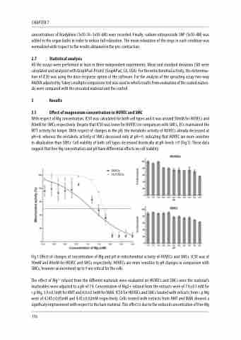
With respect of Mg concentration, IC50 was calculated for both cell types and it was around 50mM for HUVECs and 80mM for SMCs respectively. Despite that IC50 was lower for HUVECs in comparison with SMCs, ECs maintained the MTT activity for longer. With respect of changes in the pH, the metabolic activity of HUVECs already decreased at pH=8, whereas the metabolic activity of SMCs decreased only at pH=9, indicating that HUVEC are more sensitive to alkalization than SMCs. Cell viability of both cell types decreased drastically at pH-levels >9 (Fig.1). These data suggest that free Mg concentration and pH have differential effects on cell viability.
Fig.1 Effect of changes of concentration of Mg and pH in mitochondrial activity of HUVECs and SMCs. IC50 was of 50mM and 80mM for HUVEC and SMCs respectively. HUVECs are more sensitive to pH changes in comparison with SMCs, however an increment up to 9 are critical for the cells.
The effect of Mg2+ relased from the different materials were evaluated on HUVECs and SMCs once the material’s leacheables were adjusted to a pH of 7.4. Concentration of Mg2+ relased from the extracts were of 7.4±0.1 mM for c.p Mg, 3.9±0.1mM for HMT and 4.0±0.1mM for MAN. IC50 for HUVECs and SMCs treated with extracts from c.p Mg were of 4,145±0,05mM and 4.43±0,02mM respectively. Cells treated with extracts from HMT and MAN showed a significaty improvement with respect to the bare material. This effect is due to the reduced concentration of free Mg
116
