
Page 116 - Magnesium-based supports for stem cell therapy of vascular disease - Mónica Echeverry Rendón
P. 116
CHAPTER 7
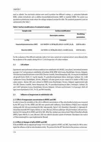
used as cathode. Two electrolyte solution were used to produce two different coatings, i.e. potassium hydroxide (KOH), sodium metasilicate and as additive hexamethylenetetramine (HMT) or mannitol (MAN). The system was operated at potentiostaic mode where the voltage remained constant for 600s. The anodized parameters used are summarized in the Table 1
Table.1 Surface modification of evaluated samples
For the evaluation of the different materials, indirect test were carried out in material extracts were obtained after the incubation of the samples during 48 h in 1.2 ml of respective cell culture medium.
2.2 Cell culture
Experiments were performed on human umbilical vein endothelial cells (HUVEC; Lonza,Basel, Switzerland) between passages 4 to 7 and growing in endothelial cell medium (ECM; RPMI 1640 (Lonza, Biowhittaker, Verviers, Belgium), 10% heat inactivated fetal bovine serum (FBS) (Thermo Scientific, Hemel Hempstead, UK), 0.06 mg/ml of endothelial cell growth factor (ECGF), 0.1 mg/mL heparin, 1% penicillin/streptomycin (Gibco, Invitrogen, Carlsbad, CA), 2 mM l-glutamine (Lonza, Biowhittaker, Verviers, Belgium). Cell were grown on pre-coated (0.1% Gelatin in PBS) tissue culture plastics. Human SMC were cultured in DMEM (Lonza Biowhittaker, Verviers, Belgium) supplemented with 10% FBS (Thermo Scientific, Hemel Hempstead, UK), 1% penicillin/streptomycin (Gibco, Invitrogen, Carlsbad, CA) and 2 mM l-glutamine (Lonza, Biowhittaker, Verviers, Belgium). Cell were used between 5 to 8 passages. Both cell types were maintained at 37oC, 5% CO2 and 98% humidity.
2.3 Influence of magnesium in endothelial cells
2.3.1 Effect of magnesium concentration in HUVEC and SMC
In order to know the sensibility of the cells to different concentrations of Mg, mitochondrial activity was measured by the MTT assay. For this, HUVEC and SMC were growth until confluency. Serial dilutions of MgCl2 were evaluated starting with 1M. Cells were incubated for 48h. Subsequently, 10μl of 3-(4,5-Dimethyl-2-thiazolyl)-2,5-diphenyl-2H- tetrazolium bromide) MTT (Sigma-Aldrich, St. Louis, Missouri, USA) was added per 100 μl of medium. Cells were incu- bated at 37oC during 4h for HUVEC and 3h for SMC. After this, medium was removed and 100μl of Dimethyl sulfoxide (DMSO, Sigma-Aldrich, St. Louis, Missouri, USA) was added to dissolve crystals of formazan. Absorbance was read at a 570nm wavelength in a spectrophotometer (Biorad).
2.3.2 Effect of changes of pH in HUVEC and SMC
ECM and DMEM with an adjusted pH (range 7.4 to 9.4) was applied to HUVEC and SMC, respectively for 48h to inves- tigate the influence of pH on mitochondrial activity. The protocol for the MTT described in the previous section was
114
