
Page 102 - Magnesium-based supports for stem cell therapy of vascular disease - Mónica Echeverry Rendón
P. 102
CHAPTER 6
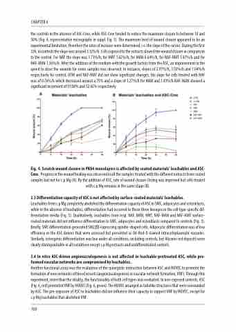
the controls in the absence of ASC-Cme, while ASC-Cme tended to reduce the maximum closure to between 10 and 50% (Fig. 4, representative micrographs in suppl. Fig. 1). The maximum level of wound closure appeared to be an experimental limitation, therefore the rates of increase were determined, i.e. the slope of the curves. During the first 32h, in controls the slope was around 3.12%/h. Cells exposed to the extracts slowed the wound closure in comparison to the control. For NAF the slope was 1.71%/h, for HMT 1.62%/h, for MAN 0.64%/h, for NAF-HMT 1.07%/h and for NAF-MAN 1.36%/h. After the addition of the medium with the growth factors from the ASC, an improvement in the speed to close the wounds for some samples was observed. In instance, slopes of 2.97%/h, 1.92%/h and 1.04%/h respectively for control, HTM and NAF-HMT did not show significant changes, the slope for cells treated with NAF was of 0.76%/h which decreased around a 75% and a slope of 1,27%/h for MAN and 1.81%/h NAF-MAN showed a significant increment of 97.08% and 32.42% respectively.
Fig. 4. Scratch wound closure in PK84 monolayers is affected by coated materials’ leachables and ASC- Cme. Progress in the wound healing was observed in all the samples treated with the different extracts from coated samples but not for c.p Mg (A). By the addition of ASC, rate of wound closure closing was improved but cells treated with c.p Mg remains in the same stage (B).
3.5 Differentiation capacity of ASC is not affected by surface-coated materials’ leachables.
Leachables from c.p Mg completely abolished the differentiation capacity of ASC to SMC, adipocytes and osteoblasts, while in the absence of leachables, differentiation had occurred to these three lineages in the cell type-specific dif- ferentiation media (Fig. 5). Qualitatively, leachables from resp. NAF, MAN, HMT, NAF-MAN and NAF-HMT surface- coated materials did not influence differentiation to SMC, adipocytes and osteoblasts compared to controls (Fig. 5). Briefly, SMC differentiation generated SM22☆-expressing spindle-shaped cells. Adipocytic differentiation was of low efficiency in the ASC donors that were assessed but presented as Oil-Red-O stained intracytoplasmatic vacuoles. Similarly, osteogenic differentiation was low under all conditions, including controls, but Alizarin-red deposits were clearly distinguishable in all conditions except c.p Mg extracts and undifferentiated controls.
3.4 In vitro ASC-driven angiovasculogenesis is not affected in leachable-pretreated ASC, while pre- formed vascular networks are compromised by leachables.
Another functional assay was the evaluation of the synergistic interaction between ASC and HUVEC to promote the formation of new networks of blood vessels (angiovasculogenesis or vascular network formation, VNF). Through this experiment, more than the vitality, the functionality of both cell types was evaluated. In non-exposed controls, ASC (Fig. 6, red) promoted VNF by HUVEC (Fig. 6, green). The HUVEC arranged as tubelike structures that were surrounded by ASC. The pre-exposure of ASC to leachables did not influence their capacity to support VNF by HUVEC, except for c.p Mg leachables that abolished VNF.
100
