
Page 101 - Magnesium-based supports for stem cell therapy of vascular disease - Mónica Echeverry Rendón
P. 101
PLASMA ELECTROLYTIC OXIDIZED MAGNESIUM ADVERSELY INFLUENCES VASCULAR CELLS TYPES BUT NOT MESENCHYMAL CELLS AND MONOCYTES
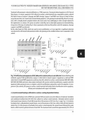
Treatment with puromycin reduced proliferation i.e. PCNA expression. Puromycin induced apoptosis in ASC from all three donors. In contrast, apoptosis of ASC adhered on surface-coated materials was comparable to plastic-cultured ASC both in terms of caspase-3 cleavage and PARP cleavage. Caspase-3 and PARP were chosen to study for these assays because these are cleaved and activated during apoptosis. Cells growing on untreated Mg showed a necrop- tosis, with a virtually absent cytoplasm which is the reason why it was challenging to collect enough material for the experiments. In contrast, ASC grown on surface-coated Mg, had no detectable apoptosis by immunoblotting, irrespective of the type of coating (Fig.3). Only in puromycin, which was the positive control for apoptosis, cleavage caspase-3 and PARP was detected.
On the other hand, for PCNA, which was used to assess proliferation, as it was expected, a significant reduction was observed in cells treated with puromycin while cells growing on the modified surfaces were comparable to the control.
6
Fig. 3 Proliferation and apoptosis of ASC adhered to coated surfaces is not affected. Immunoblotting with antibodies against PCNA (proliferation), caspase-3, which detects cleaved caspase-3 (apoptosis) as well and PARP, which detects cleaved PARP (apoptosis) as well was done ASC cultured on surface-coated materials (NAF, HMT, MAN, NAF/HMT, NAF/MAN) and tissue culture plastic (untreated cells, positive control for proliferation, negative control for apoptosis) while puromycin serves as negative control for proliferation and positive control for apoptosis. In com- parison to the controls, ASC grown on surface-coated materials had similar levels of proliferation and no apoptosis. All data were normalized with respect to β-tubulin.
3.2 Scratch wound healing is affected in a surface-coating-dependent fashion.
Scratch wound healing in vitro of PK84 over a period of 48h was used to assess the influence of materials’ leachables and conditioned medium from ASC (ASC-Cme) cultured on surface-coated materials. Untreated controls reached total wound closure between 24 and 48h irrespective of the presence of ASC-Cme (Fig. 4, suppl. Fig. 1). In contrast, leachables from c.p Mg abolished wound closure, even in the presence of ASC-Cme, while at 48h cells had started to detach (Fig. 4). In the presence of leachables from surface-coated materials, the wound closure followed first order kinetics which reached a plateau between 24 and 48h. The maximum levels varied from approximately 20 to 70% of
99
