
Page 276 - Demo
P. 276
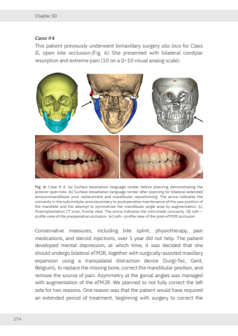
Chapter 10274Case #4This patient previously underwent bimaxillary surgery alio loco for Class II, open bite occlusion.(Fig. 6) She presented with bilateral condylar resorption and extreme pain (10 on a 0–10 visual analog scale). Fig. 6: Case # 4. (a) Surface tesselation language render before planning demonstrating the anterior open bite. (b) Surface tessellation language render after planning for bilateral extended temporomandibular joint replacement and mandibular repositioning. The arrow indicates the concavity in the subcondylar area secondary to postoperative maintenance of the yaw position of the mandible and the attempt to symmetrize the mandibular angle area by augmentation. (c) Postimplantation CT scan, frontal view. The arrow indicates the mid-cheek concavity. (d) Left — profile view of the preoperative occlusion. (e) Left— profile view of the post‑eTMJR occlusionConservative measures, including bite splint, physiotherapy, pain medications, and steroid injections, over 1 year did not help. The patient developed mental depression, at which time, it was decided that she should undergo bilateral eTMJR, together with surgically-assisted maxillary expansion using a transpalatal distraction device (Surgi-Tec, Gent, Belgium), to replace the missing bone, correct the mandibular position, and remove the source of pain. Asymmetry at the gonial angles was managed with augmentation of the eTMJR. We planned to not fully correct the left side for two reasons. One reason was that the patient would have required an extended period of treatment, beginning with surgery to correct the Nikolas de Meurechy NW.indd 274 05-06-2024 10:15
