
Page 273 - Demo
P. 273
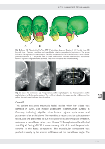
e-TMJ replacement: Clinical analysis27110Fig. 2: Case #1. Planning in ProPlan CMF (Materialize, Leuven, Belgium). (A) Frontal view. (B) Frontal view. Planned maxillary and mandibular rotation repositioning osteotomy. The arrow indicates the sagittal split osteotomy on the side contralateral to the extended temporomandibular joint replacement. (C) Left profile view. (D) Left profile view. Planned maxillary and mandibular rotation repositioning osteotomy planned. The arrow indicates the coronoidotomyFig. 3: Case #1 continued. (a) Preoperative profile cephalogram. (b) Postoperative profile cephalogram. (c) Orthopantomogram. The red line indicates the upper dental midline, and the yellow line indicates the lower dental midlineCase #2This patient sustained traumatic facial injuries when her village was bombed in 2007. She initially underwent reconstruction surgery in Germany, including polyether ether ketone zygoma replacement and placement of an artificial eye. The mandibular reconstruction subsequently failed, and she presented to our institution with a chronic plate infection, malunion, a mandibular defect, and fibrous TMJ ankylosis on the affected side.(Fig. 4) During eTMJR, it was extremely difficult to seat the prosthetic condyle in the fossa component. The mandibular component was pushed inwardly by the scarred soft tissues at the mandibular angle. The Nikolas de Meurechy NW.indd 271 05-06-2024 10:15
