
Page 229 - Demo
P. 229
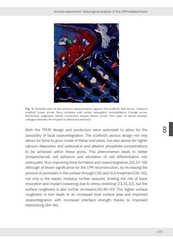
Animal experiment: Histological analysis of the LPM reattachment2278Fig. 5: Detailed view of the enthesis approximation against the scaffold. Red arro Titanium scaffold Green arro Bony enthesis with active osteogenic remodellation Orange arro Storiformly organized, dense connective tissues Yellow arro Thin layer of dense lamellar collagen between the implant scaffold and enthesisBoth the TMJR design and production were optimized to allow for the possibility of local osseointegration. The scaffold’s porous design not only allows for bone to grow inside of these void areas, but also allows for higher calcium deposition and osteocalcin and alkaline phosphate concentrations to be achieved within these pores. This phenomenon leads to better (mesenchymal) cell adhesion and elicitation of cell differentiation into osteocytes, thus improving bone formation and osseointegration.(20,33–38) Although of lesser significance for the LPM reconstruction, by increasing the amount of porosities in the surface through CAD and SLA-treatment(28–30), not only is the elastic modulus further reduced, limiting the risk of bone resorption and implant loosening due to stress shielding (23,31,32), but the surface roughness is also further increased.(30,40–43) This higher surface roughness in turn leads to an increased total surface area and improved osseointegration with increased interface strength thanks to improved interlocking.(44–46) Nikolas de Meurechy NW.indd 227 05-06-2024 10:14
