
Page 221 - Demo
P. 221
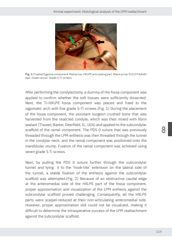
Animal experiment: Histological analysis of the LPM reattachment2198Fig. 1: Fixated Zygoma component. Red arro HXLPE articulating part. Black arro ELI23Ti6Al4V part. Green arro Grade 5-Ti screwsAfter performing the condylectomy, a dummy of the fossa component was applied to confirm whether the soft tissues were sufficiently dissected. Next, the Ti-HXLPE fossa component was placed and fixed to the zygomatic arch with five grade 5-Ti screws.(Fig. 1) During the placement of the fossa component, the assistant surgeon crushed bone that was harvested from the resected condyle, which was then mixed with fibrin sealant (Tisseel; Baxter, Deerfield, IL, USA) and applied to the subcondylar scaffold of the ramal component. The PDS 0 suture that was previously threaded through the LPM enthesis was then threaded through the tunnel in the condylar neck, and the ramal component was positioned onto the mandibular stump. Fixation of the ramal component was achieved using seven grade 5 Ti screws.Next, by pulling the PDS 0 suture further through the subcondylar tunnel and tying it to the ‘hook-like’ extension on the lateral side of the tunnel, a stable fixation of the enthesis against the subcondylar scaffold was attempted.(Fig. 2) Because of an obstructive caudal edge at the anteromedial side of the HXLPE part of the fossa component, proper approximation and visualization of the LPM enthesis against the subcondylar scaffold proved challenging. Consequently, all the HXLPE parts were scalpel-reduced at their non-articulating anteromedial side. However, proper approximation still could not be visualized, making it difficult to determine the intraoperative success of the LPM reattachment against the subcondylar scaffold.Nikolas de Meurechy NW.indd 219 05-06-2024 10:14
