
Page 196 - Demo
P. 196
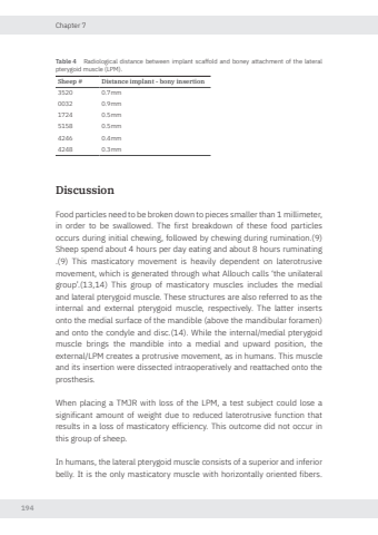
Chapter 7194Table 4 Radiological distance between implant scaffold and boney attachment of the lateral pterygoid muscle (LPM).Sheep # Distance implant - bony insertion3520 0.7mm0032 0.9mm1724 0.5mm5158 0.5mm4246 0.4mm4248 0.3mmDiscussion Food particles need to be broken down to pieces smaller than 1 millimeter, in order to be swallowed. The first breakdown of these food particles occurs during initial chewing, followed by chewing during rumination.(9) Sheep spend about 4 hours per day eating and about 8 hours ruminating .(9) This masticatory movement is heavily dependent on laterotrusive movement, which is generated through what Allouch calls ‘the unilateral group’.(13,14) This group of masticatory muscles includes the medial and lateral pterygoid muscle. These structures are also referred to as the internal and external pterygoid muscle, respectively. The latter inserts onto the medial surface of the mandible (above the mandibular foramen) and onto the condyle and disc.(14). While the internal/medial pterygoid muscle brings the mandible into a medial and upward position, the external/LPM creates a protrusive movement, as in humans. This muscle and its insertion were dissected intraoperatively and reattached onto the prosthesis.When placing a TMJR with loss of the LPM, a test subject could lose a significant amount of weight due to reduced laterotrusive function that results in a loss of masticatory efficiency. This outcome did not occur in this group of sheep. In humans, the lateral pterygoid muscle consists of a superior and inferior belly. It is the only masticatory muscle with horizontally oriented fibers.Nikolas de Meurechy NW.indd 194 05-06-2024 10:14
