
Page 195 - Demo
P. 195
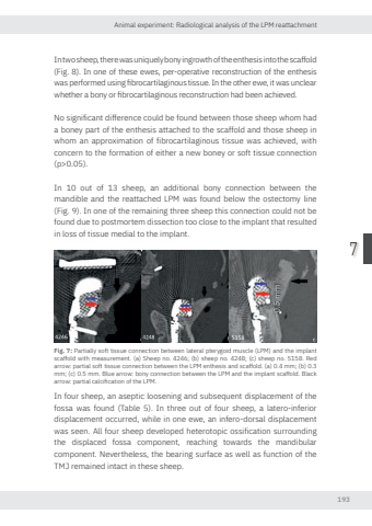
Animal experiment: Radiological analysis of the LPM reattachment1937In two sheep, there was uniquely bony ingrowth of the enthesis into the scaffold (Fig. 8). In one of these ewes, per-operative reconstruction of the enthesis was performed using fibrocartilaginous tissue. In the other ewe, it was unclear whether a bony or fibrocartilaginous reconstruction had been achieved. No significant difference could be found between those sheep whom had a boney part of the enthesis attached to the scaffold and those sheep in whom an approximation of fibrocartilaginous tissue was achieved, with concern to the formation of either a new boney or soft tissue connection (p>0.05).In 10 out of 13 sheep, an additional bony connection between the mandible and the reattached LPM was found below the ostectomy line (Fig. 9). In one of the remaining three sheep this connection could not be found due to postmortem dissection too close to the implant that resulted in loss of tissue medial to the implant.Fig. 7: Partially soft tissue connection between lateral pterygoid muscle (LPM) and the implant scaffold with measurement. (a) Sheep no. 4246; (b) sheep no. 4248; (c) sheep no. 5158. Red arro partial soft tissue connection between the LPM enthesis and scaffold. (a) 0.4 mm; (b) 0.3 mm; (c) 0.5 mm. Blue arro bony connection between the LPM and the implant scaffold. Black arro partial calcification of the LPM.In four sheep, an aseptic loosening and subsequent displacement of the fossa was found (Table 5). In three out of four sheep, a latero-inferior displacement occurred, while in one ewe, an infero-dorsal displacement was seen. All four sheep developed heterotopic ossification surrounding the displaced fossa component, reaching towards the mandibular component. Nevertheless, the bearing surface as well as function of the TMJ remained intact in these sheep.Nikolas de Meurechy NW.indd 193 05-06-2024 10:14
