
Page 168 - Demo
P. 168
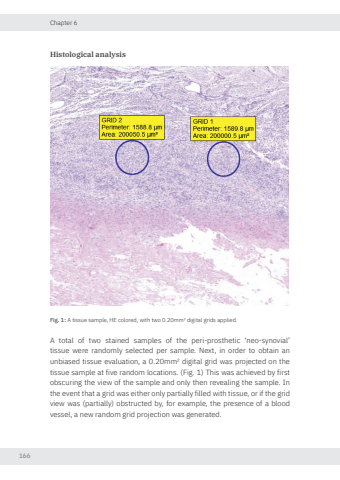
Chapter 6166Histological analysisFig. 1: A tissue sample, HE colored, with two 0.20mm² digital grids applied.A total of two stained samples of the peri-prosthetic ‘neo-synovial’ tissue were randomly selected per sample. Next, in order to obtain an unbiased tissue evaluation, a 0.20mm² digital grid was projected on the tissue sample at five random locations. (Fig. 1) This was achieved by first obscuring the view of the sample and only then revealing the sample. In the event that a grid was either only partially filled with tissue, or if the grid view was (partially) obstructed by, for example, the presence of a blood vessel, a new random grid projection was generated. Nikolas de Meurechy NW.indd 166 05-06-2024 10:14
