
Page 42 - Development of Functional Scaffolds for Bone Tissue Engineering Using 3D-Bioprinting of Cells and Biomaterials - Yasaman Zamani
P. 42
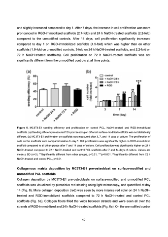
and slightly increased compared to day 1. After 7 days, the increase in cell proliferation was more pronounced in RGD-immobilized scaffolds (2.7-fold) and 24 h NaOH-treated scaffolds (2.2-fold) compared to the unmodified controls. After 14 days, cell proliferation significantly increased compared to day 1 on RGD-immobilized scaffolds (4.5-fold) which was higher than on other scaffolds (1.9-fold on unmodified controls, 3-fold on 24 h NaOH-treated scaffolds, and 2.2-fold on 72 h NaOH-treated scaffolds). Cell proliferation on 72 h NaOH-treated scaffolds was not significantly different from the unmodified controls at all time points.
Figure 5. MC3T3-E1 seeding efficiency and proliferation on control PCL, NaOH-treated, and RGD-immobilized scaffolds. (a) Seeding efficiency measured 12 h post seeding on different surface-modified scaffolds was not statistically different. (b) MC3T3-E1 proliferation on scaffolds was measured after 3, 7, and 14 days of culture. The proliferation of cells on the scaffolds were compared relative to day 1. Cell proliferation was significantly higher on RGD-immobilized scaffold compared to all other groups after 7 and 14 days of culture. Cell proliferation was significantly higher on 24 h NaOH-treated compared to 72 h NaOH-treated and control PCL scaffolds after 7 and 14 days of culture. Values are mean ± SD (n=3). **Significantly different from other groups, p<0.01, ***p<0.001, ##significantly different from 72 h NaOH-treated and control PCL, p<0.01.
Collagenous matrix deposition by MC3T3-E1 pre-osteoblast on surface-modified and unmodified PCL scaffolds
Collagen deposition by MC3T3-E1 pre-osteoblasts on surface-modified and unmodified PCL scaffolds was visualized by picrosirius red staining using light microscopy, and quantified at day 14 (Fig. 6). More collagen deposition (red) was seen by more intense red color on 24 h NaOH- treated and RGD-immobilized scaffolds compared to 72 h NaOH-treated and control PCL scaffolds (Fig. 6a). Collagen fibers filled the voids between strands and were seen all over the strands of RGD-immobilized and 24 h NaOH-treated scaffolds (Fig. 6a). On the unmodified control
 40
