
Page 41 - Development of Functional Scaffolds for Bone Tissue Engineering Using 3D-Bioprinting of Cells and Biomaterials - Yasaman Zamani
P. 41
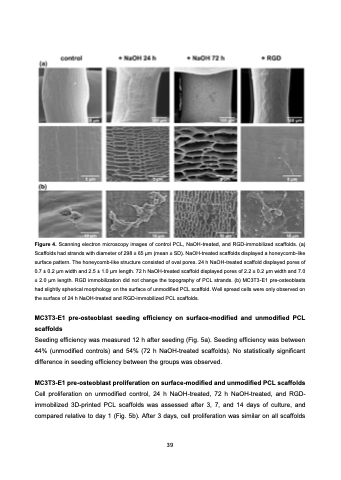
 Figure 4. Scanning electron microscopy images of control PCL, NaOH-treated, and RGD-immobilized scaffolds. (a) Scaffolds had strands with diameter of 298 ± 65 μm (mean ± SD). NaOH-treated scaffolds displayed a honeycomb-like surface pattern. The honeycomb-like structure consisted of oval pores. 24 h NaOH-treated scaffold displayed pores of 0.7 ± 0.2 μm width and 2.5 ± 1.0 μm length. 72 h NaOH-treated scaffold displayed pores of 2.2 ± 0.2 μm width and 7.0 ± 2.0 μm length. RGD immobilization did not change the topography of PCL strands. (b) MC3T3-E1 pre-osteoblasts had slightly spherical morphology on the surface of unmodified PCL scaffold. Well spread cells were only observed on the surface of 24 h NaOH-treated and RGD-immobilized PCL scaffolds.
MC3T3-E1 pre-osteoblast seeding efficiency on surface-modified and unmodified PCL scaffolds
Seeding efficiency was measured 12 h after seeding (Fig. 5a). Seeding efficiency was between 44% (unmodified controls) and 54% (72 h NaOH-treated scaffolds). No statistically significant difference in seeding efficiency between the groups was observed.
MC3T3-E1 pre-osteoblast proliferation on surface-modified and unmodified PCL scaffolds
Cell proliferation on unmodified control, 24 h NaOH-treated, 72 h NaOH-treated, and RGD- immobilized 3D-printed PCL scaffolds was assessed after 3, 7, and 14 days of culture, and compared relative to day 1 (Fig. 5b). After 3 days, cell proliferation was similar on all scaffolds
39
