
Page 78 - Fluorescence-guided cancer surgery
P. 78
76
Chapter 5
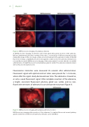
Figure 2. NIR uorescence imaging of a pituitary adenoma
A. NIR uorescence imaging of adenoma and normal gland after partial resection of the adenoma. ICG was administered just before opening of the dura, a second bolus of ICG was planned just after making this image. In the color image, a dark red colored remnant of the adenoma is visible. In the NIR uorescence image, a slightly uorescent normal gland is visible (circle). The adenoma remnant does not stain uorescent. B. NIR uorescence imaging of the same patient 60 seconds after the second ICG administration. A bright uorescent normal pituitary gland is visible. The adenoma remnant remains still non- uorescent (dashed circle).
Fluorescence intensities were measured 45 seconds after administration. Fluorescent signal with optimal contrast ratios were present for 1-2 minutes, where after the signal slowly decreased over time. The adenomas showed no, or only a weak uorescent signal. After complete resection of the adenoma, a bright, consistent uorescent pituitary gland was visible, and no non- uorescent remnants of adenoma tissue were present anymore (Figure 3).
Figure 3. NIR uorescence imaging after pituitary adenoma resection
NIR uorescence imaging after resection of the adenoma. A complete uorescent normal pituitary gland is visible. No non- uorescent adenoma remnants can be identi ed.
