
Page 77 - Fluorescence-guided cancer surgery
P. 77
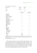
Table 1. Patient and tumor characteristics
Characteristic
Age
Gender
Median
50
N (n = 10)
Range
28 - 74
%
Imaging in pituitary surgery 75
M 440 F 660
Preoperative NN
7 10 2 0 1 0
Postoperative
Visual de cits
No de cits
Minor de cits
Mild bitemporal de cits
Cranial nerve de cits
None 88
Total N.III OS
Mild N.III OD
Moderate N.III, slight N.VI OD Minor N.VI OD
Pituitary Function
No de cits Hypogonadism Hypothyriodism
Hypersecretion
None
ACTH 40 PRL 10 GH and PRL 10
Pathology Biochemical
NFMA 44
Cushing’s disease Acromegaly Prolactinoma
IHC
GH+; PRL + ACTH+ LH+ Null-cell
4 4 1 1 1 1
N/A 2 N/A 5 N/A 1 N/A 2
1 0 0 1* 1 0 0 1*
6 8 3 1 1 1
4 10
Abbreviations: ACTH, adrenocorticotropic hormone; DI, Diabetes Insipidus; GH, Growth hormone; IHC, immunohistochemistry; LH, Luteinizing hormone; N/A, not applicable; OD, oculus dextra; OS, oculus sinistra; PRL, prolactin.
* Recovered completely after 6 months
After opening of the dura, the normal pituitary gland showed a more intense  uorescent signal than the adenoma in nine of the ten patients. In one patient intercavernous sinus venous bleeding prevented assessment of the  uorescent signal. Figure 2 shows images of the  uorescent signal in adenoma and normal gland during resection. In general, during resection, a bright  uorescent normal gland became visible after ICG administration.
