
Page 76 - Fluorescence-guided cancer surgery
P. 76
74
Chapter 5
RESULTS
In the inclusion period 58 patients underwent a transnasal transsphenoid operation. Fourteen patients had a di erent pathology than adenoma. Six patients had been operated before. One patient was allergic to iodine. Ten patients refused participation in the study for a variety of reasons.
The light cable was temporarily broken leading to an inclusions stop for 4 weeks and subsequently exclusion of 7 patients. From the remaining 20 patients always the rst patient of the two cases scheduled for surgery that day was included, leading to 10 included patients.
Median age was 50 years, ranging between 28 and 74. Six patients were female. Patients had either clinical features and imaging consistent with Non- Functioning macro-Adenoma (NFMA) (N=4), or were biochemically diagnosed with a functional adenoma (Cushing’s disease (N=4), Acromegaly (N=1) and Prolactinoma (N=1)). Patient and tumor characteristics are shown in Table 1. All resected lesions were histologically proven to be a pituitary adenoma.
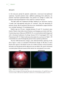
The rst NIR uorescence images were obtained before opening of the dura. Figure 1 shows an example of uorescent images of the dura. A clear uorescent signal in the blood vessels was visible, however no discrimination between normal gland and the adenoma was possible at this point. Enhanced vascularity due to dural invasion of the tumor was not present in our series.
Figure 1. NIR uorescence imaging of the dura
NIR uorescence imaging of the unopened dura mater after administration of 5 mg ICG. Fluorescent vessels are visible on the dura. No identi cation of adenoma or normal pituitary gland is observed.
