
Page 66 - Fluorescence-guided cancer surgery
P. 66
64
Chapter 4
Figure 3. Fluorescent guidance during resection
NIR uorescence imaging was used as guidance during resection of uveal liver metastases. Resection
was performed using an Endo GIATM 45 reload stapler (Covidien) (asterisk).
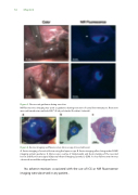
Figure 4. Ex vivo imaging and uorescence microscopy of resected lesion
A. Ex vivo imaging of resected lesion using the laparoscope. B. Ex vivo imaging after slicing under FLARE imaging system guidance. C. Microscopic overlay of Hematoxylin and Eosin staining of the resected lesion and uorescent signal (Odyssey Infrared Imaging System, LI-COR). A clear uorescent rim was observed around the malignant lesion.
No adverse reactions associated with the use of ICG or NIR uorescence imaging were observed in any patient.
