
Page 64 - Fluorescence-guided cancer surgery
P. 64
62
Chapter 4
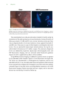
Figure 1. Identi cation of liver metastases
Multiple uveal liver metastases, identi ed by inspection and NIR uorescence imaging as a typical uorescent rim (arrows) around each lesion, 24 h after intravenous injection of 10 mg ICG. Open arrows indicate the preoperative known metastasis.
The second patient was a 66-year-old woman treated 22 months earlier by enucleation of the right eye because of uveal melanoma. During 20 months of follow-up using US, CT, MRI and PET scans, no liver metastases were seen. Now she was diagnosed with a 12 mm liver metastasis in liver segment III on both CT and MRI scans. There were no signs of other hepatic or extrahepatic metastatic lesions. She was planned for laparoscopic metastasectomy. Ten mg ICG was administered intravenously 24 h before surgery. The previously diagnosed lesion was detected by inspection, laparoscopic ultrasonography (LUS), and NIR uorescence imaging, and was removed under uorescent guidance (Figures 2a and 3; supplementary video’s, online available).
Using NIR uorescence imaging, an additional suspicious lesion in segment III was identi ed, which showed a typical 1.5 mm uorescent rim (Figure 2b). This lesion was characterized as a hemangioma by inspection, and was not identi ed with LUS. It was resected under uorescent guidance. Both removed lesions proved to be malignant by histological assessment and were radically removed (Figure 4). No other lesions were identi ed by inspection, LUS or NIR uorescence imaging. Patient recovered well from surgery, and no recurrent disease was seen during 15 months of follow-up.
