
Page 65 - Fluorescence-guided cancer surgery
P. 65
Laparoscopic imaging of hepatic uveal melanoma metastases 63
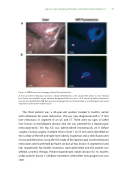
Figure 2. NIR uorescence imaging of uveal liver metastases
A. An uveal liver metastasis (arrow) is clearly identi ed by a rim around the tumor in vivo. Normal liver tissue (arrowhead) shows minimal background uorescence. B. A small super cial metastasis (arrow) was identi ed by NIR uorescence imaging, but was characterized as a hemangioma by visual inspection and was not visible by LUS.
The third patient was a 65-year-old woman treated 6 months earlier with ruthenium for uveal melanoma. She was now diagnosed with a 17 mm liver metastasis in segment IV on US and CT. There were no signs of other liver lesions or extrahepatic disease and she was planned for a laparoscopic metastasectomy. Ten mg ICG was administered intravenously 24 h before surgery. During surgery, multiple lesions from 1 to 10 mm were identi ed on the surface of the left and right liver lobe by inspection and a clear uorescent rim around the tumor using the ICG mode of the laparoscope. Uveal melanoma metastases were con rmed by frozen section of two lesions in segments III and IVb, respectively. No further resections were performed and the patient was o ered systemic therapy. Patient experienced stable disease for 16 months under protein kinase C inhibitor treatment, whereafter new progression was seen.
