
Page 31 - Fluorescence-guided cancer surgery
P. 31
SLN detection in gastric cancer 29
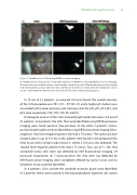
Figure 1. Identi cation of SLNs using NIR uorescence imaging
A. Identi cation of SLN (arrow) 15 min after injection of ICG:Nanocoll using NIR uorescence imaging. The injection site around the tumor is indicated by a dashed arrow. B. Patient with tumor-positive lymph node (indicated by arrow). Injection sites and uorescent SLN are clearly detected. Lymphatic vessels are also visible between the injection site and SLN. The SLN is marked using sutures.
In 19 out of 21 patients, an accurate SLN was found. The overall accuracy of the SLN procedure was 90% (70 – 99 95% CI) and a higher pT-stadium was associated with a lower accuracy rate. Accuracy rates for pTx, pT1, pT2, pT3, and pT4 were respectively 100, 100, 100, 90, and 0%.
Histological analysis of the SLNs showed lymph node metastases in 8 out of 21 patients. In 6 patients, the SLNs that were identi ed using NIR uorescence imaging were tumor-positive (true positive). In the other 2 patients, tumor- positive lymph nodes were not identi ed using NIR uorescence imaging (false- negative). One false-negative patient (#2) had a T4 tumor. The tumor positive lymph nodes (3 out of 33 LNs) in this patient were found in the peripancreatic fatty tissue and in lymph node station 3, where a SLN was also detected. The second false-negative patient (#10) had a T3 tumor. Four out of 11 LNs that contained tumor cells were not detected by NIR uorescence imaging. Of particular importance, all 7 tumor-positive LNs that were not detected by NIR uorescence imaging were completely e aced by tumor tissue and no lymphatic tissue could be identi ed.
In 8 patients, SLNs outside the standard resection plane were identi ed. In 4 patients these were located in the hepatoduodenal ligament (LN station
