
Page 29 - Fluorescence-guided cancer surgery
P. 29
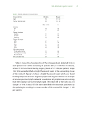
Table 1. Patient and tumor characteristics
Characteristic
Age
Tumor size (in mm)
Gender
Median
64 35
N (n = 26)
Range
30 - 82 10 – 90
%
M 1973 F 727
Tumor location
Cardia Corpus Antrum
Tumor pT stage
8 31
6 23 12 46
pTx 28 pT1 519 pT2 519 pT3 10 39 pT4 415
Type of resection
Total gastrectomy Partial gastrectomy No resection
Preoperative CTx
11 42 14 54 1 4
23 88
SLN detection in gastric cancer 27
Table 2 shows the characteristics of the intraoperatively detected SLNs in each patient. In 21 of the remaining 22 patients (95%, 77–100 95% CI interval), at least 1 SLN was found during surgery (mean of 3.1 SLNs per patient; range 1-6). SLNs were identi ed as bright  uorescent spots in the surrounding tissue of the stomach. Figure 1A shows a bright  uorescent spot, which was found histologically to be a tumor-negative lymph node. Figure 1B shows an example of a tumor-positive lymph node and visualization of lymphatic vessels running from the injection site to the lymph node. The mean SBR of the SLNs was 4.4 (range 1.4–19.8). In total, 533 LNs were identi ed in the resection specimens by the pathologist, resulting in a mean number of 24 resected LNs (range 11 - 44) per patient.
