
Page 32 - Fluorescence-guided cancer surgery
P. 32
30
Chapter 2
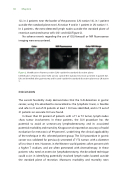
12), in 2 patients near the border of the pancreas (LN station 14), in 1 patient outside the standard plane near LN station 9 and in 1 patient in LN station 11. In 2 patients, the extra-detected lymph nodes outside the standard plane of resection contained tumor cells (#21 and #22)(Figure 2).
No adverse events regarding the use of ICG:Nanocoll or NIR uorescence imaging were encountered.
Figure 2. Identi cation of tumor-positive SLNs outside the standard dissection planes
Identi cation of tumor-positive SLNs (arrow) outside the standard resection specimen in patient #21. LNs are identi ed after gastrectomy and located outside the standard dissection plane near LN station 9.
DISCUSSION
The current feasibility study demonstrates that the SLN detection in gastric cancer, using ICG adsorbed to nanocolloid as the lymphatic tracer, is feasible and safe. In 21 out of 22 patients at least 1 SLN was identi ed, and in 19 out of 21 patients an accurate SLN was found.
In fewer than 50 percent of patients with a T1 or T2 tumor, lymph nodes show tumor involvement. In these patients, the SLN procedure has the potential to avoid an unnecessary lymphadenectomy, and its associated potential morbidity and mortality. Kitagawa et al reported an accuracy of nodal evaluation for metastasis of 99 percent7, underlining the clinical applicability of the technique in this selected patient group. The SLN procedure in gastric cancer was validated for previously untreated cT1-T2 tumors with a diameter of less than 4 mm. However, in the Western world patients often present with a higher T stadium, and are often pretreated with chemotherapy. In these patients who need an extensive lymphadenectomy, the described technique could assist in identifying potentially involved lymph nodes located outside the standard plane of resection. Moreover, morbidity and mortality rates
