
Page 171 - Fluorescence-guided cancer surgery
P. 171
Clinical translation of OTL38 in ovarian cancer 169
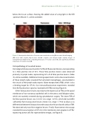
below the tissue surface, showing the added value of using light in the NIR spectrum (Movie S1, online available).
Figure 3. Intraoperative detection of ovarian cancer metastases using uorescence-based imaging
A-B. Color (left column), uorescence (middle column), and merged (right column) images of retroperitoneal lymph nodes containing metastases of ovarian cancer (A) and super cial peritoneal metastases of ovarian cancer (B).
Histopathology of resected lesions
No malignant disease was found in 21 of the 83 uorescent lesions, corresponding to a false positive rate of 23%. These false positive lesions were observed primarily in lymph nodes, representing 52% of all false positive lesions (Table S5, online available). Additional staining experiments and a closer examination of these lymph nodes revealed that activated macrophages, accumulated in the sinuses of the lymph node, express folate receptor beta (FRβ), which is also a binding target for OTL38. Our immuno uorescence experiments revealed that the uorescence signal co-localized with FRβ staining (Figure 4).
Other false positive results arose due to the expression of FRα on the apical membrane of non-cancerous epithelial cells in the uterus and fallopian tubes, which are routinely resected during cytoreductive surgery. The mean TBR of the false positive lesions was 5.4 (SD: 2.0, range: 1.8-9.3), which did not di er su ciently from true positive lesions (mean: 4.4, range: 1.7-9.8) to allow us to di erentiate between false positive and true positive lesions based solely on TBR. We observed only two false negative lesions. Finally, uorescence microscopy revealed the accumulation of OTL38 in the membrane and cytoplasm of FRα- expressing tumor cells (for representative images, see Figure 5).
