
Page 172 - Fluorescence-guided cancer surgery
P. 172
170
Chapter 10
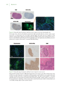
Figure 4. Histopathological evaluation and uorescence signal of a false positive lymph node
The images show a uorescent lymph node (bottom right panel) that did not contain ovarian cancer metastases on hematoxylin and eosin (H&E) staining (upper left panel) and was negative for FRα immuno staining (upper right panel). Immunostaining for FRβ showed positive staining in the sinuses, but not in the follicles (lower left panel). The magni ed image at the right shows that the uorescence pattern was localized to the sinuses, consistent with FRβ staining.
Figure 5. Histopathological evaluation and uorescence signal in ovarian cancer
Representative samples from two di erent patients. Fluorescence microscopy (left column) shows clear membranous and cytoplasmic accumulation of OTL38 in tumor cells. The uorescence signal is indicated in green, and the nuclei were counterstained with DAPI (blue). The uorescence pattern is consistent with FRα expression measured using immunohistochemistry (middle column), which corresponds to the anatomical site containing a serous ovarian adenocarcinoma metastasis visible on hematoxylin and eosin (H&E) staining (right column, dashed outline).
