
Page 176 - Assessing right ventricular function and the pulmonary circulation in pulmonary hypertension Onno Anthonius Spruijt
P. 176
10
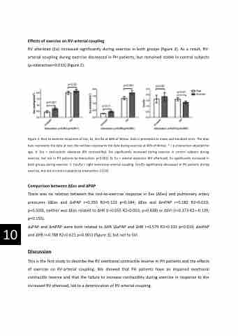
Effects of exercise on RV-arterial coupling
RV afterload (Ea) increased significantly during exercise in both groups (figure 2). As a result, RV- arterial coupling during exercise decreased in PH patients, but remained stable in control subjects (p-interaction=0.013) (figure 2).
Figure 2: Rest-to-exercise responses of Ees, Ea, Ees/Ea at 40% of Wmax. Data is presented as mean and standard error. The blue bars represents the data at rest, the red bars represents the data during exercise at 40% of Wmax. * = p-interaction adjusted for age. A: Ees = end-systolic elastance (RV contractility). Ees significantly increased during exercise in control subjects during exercise, but not in PH patients (p-interaction: p<0.001). B: Ea = arterial elastance (RV afterload). Ea significantly increased in both groups during exercise. C: Ees/Ea = right ventricular-arterial coupling. Ees/Ea significantly decreased in PH patients during exercise, but not in control subjects (p-interaction: 0.013).
Comparison between ΔEes and ΔPAP
There was no relation between the rest-to-exercise response in Ees (ΔEes) and pulmonary artery pressures (ΔEes and ΔsPAP r=0.350 R2=0.123 p=0.184; ΔEes and ΔmPAP r=0.182 R2=0.033; p=0.500), neither was ΔEes related to ΔHR (r=0.055 R2=0.003; p=0.838) or ΔSVI (r=0.373 R2=-0.139; p=0.155).
ΔsPAP and ΔmPAP were both related to ΔHR (ΔsPAP and ΔHR r=0.579 R2=0.335 p=0.019; ΔmPAP and ΔHR r=0.788 R2=0.621 p<0.001) (figure 3), but not to SVI.
Discussion
This is the first study to describe the RV exertional contractile reserve in PH patients and the effects of exercise on RV-arterial coupling. We showed that PH patients have an impaired exertional contractile reserve and that the failure to increase contractility during exercise in response to the increased RV afterload, led to a deterioration of RV-arterial coupling.
