
Page 160 - Assessing right ventricular function and the pulmonary circulation in pulmonary hypertension Onno Anthonius Spruijt
P. 160
9
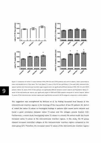
Figure 5: Comparison of native T1-values between IPAH, PAH-SSc and CTEPH patients and control subjects. Data is presented as mean and standard error of the mean. The Y-axis Native T1-values of the RV free wall (blue), LV free wall (red), interventricular septum (yellow) and interventricular insertion regions (green) were not significantly different between IPAH, PAH-SSc and CTEPH patients. Native T1-values of the LV free wall was not significantly different between control subjects and PH patients. Native T1- values of the interventricular septum was significantly higher in IPAH and CTEPH patients compared to control subjects. Native T1-values of the interventricular insertion regions were significantly increased in all PH categories compared to control subjects.
This suggestion was strengthened by McCann et al. by finding increased focal fibrosis at the interventricular insertion regions in the histology of the myocardium of two PH patients [4]. Bull et al. linked the native T1-values to histological findings in patients with severe aortic stenosis and found a good correlation between native T1-values and the collagen volume fraction [12]. Furthermore, a recent study investigating native T1-values in a chronic PH animal model also found increased native T1-values at the interventricular insertion regions. In this study, the PH group showed increased interstitial collagen at the interventricular insertion regions compared to the sham group [27]. Therefore, the increased native T1-values of the interventricular insertion regions
