
Page 39 - scheppingen
P. 39
NOVEL HISTOPATHOLOGICAL PATTERNS IN CORTICAL TUBERS IN TSC
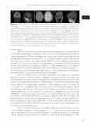
Figure 5 Clinical implications. A. Representative MRI of a patient with a histological type A tuber (histology is shown in Fig. 1B) characterized by a hyperintense lesion parasaggital in the right fron- tal lobe (indicated by the arrow head) on T2 weighted and fluid-attenuated inversion recovery (FLAIR) and a hypointense signal in the same area on volumetric T1 images. B. MRI of a histo- logical type B tuber (histology is shown in Fig. 1C) with FCD-like features in the left postcentral/ parietal region, showing as a hyperintense lesion on FLAIR image and a hypointense signal on 3DT1. FCD features are recognized by thickened cortex,blurring of gray and white matter junction and a transmantal sign. C. MRI of a histological type C tuber (histology is shown in Fig. 1D) with a large calcification, characterized by deep hypointense signal with surrounding heterogenous hy- perintense signal on T2 weighted and FLAIR images and a hypointense signal in the white matter on 3DT1. The above described FCD features are seen here as well.
cortical tubers.
Recently, two new classification schemes were presented by task forces of the
International League Against Epilepsy (ILAE). In 2011, FCD, the most common cause of intractable epilepsy in children, was addressed 17, and in 2013 a novel classification scheme for hippocampal sclerosis was published 18. These schemes, however, were established to distinguish distinct entities considering clearly different clinical etiologies. Nevertheless, the new FCD classification scheme has already proven to be more reliable with regard to inter-rater variability than previous schemes 19, 20. Most importantly, the first reports on possible clinical value for prediction of surgical outcome have been published 21, 22. In these studies, seizure-freedom was dependent on accurate definition of the epilepto- genic zone and the subsequent extent of surgical resection 21, 22.
Until now cortical tubers have been neglected in this respect. In an attempt to meet the current need of a better histological assessment we have identified three distinct patterns of cortical tubers. Consequently, we were able to show that our tuber patterns are recognizable by different neuropathologists and therefore reasonably appli- cable in different neuropathology laboratories. In our endeavor to identify variants of TSC lesions on a histological level and to stay within the frame of diagnostic useful- ness and high accessibility we chose a panel of antibodies that has been established and widely-used previously and subsequently used statistic modeling to asses quantitative differences 23. The normal expression pattern of the selected markers has been also evaluated in post-mortem control tissue. However, one limitation of this study is the availability of brain tissue within the age range of the TSC subjects. An ideal experimental design (including also ages ranging between 1 and 18 year of age) is difficult to achieve and representative material from patients without any significant brain pathology is not available at all developmental ages.
There are only a few studies which report highly advanced methods of quanti- tative histology on epilepsy surgery specimens 24, 25, 26, 14. Most of these addressed very specific pathological features to show differences in expression patterns and therefore a lot of the data were generated via a region of interest (ROI) based approach. So far,
37
two
