
Page 38 - scheppingen
P. 38
two
36
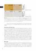
Figure 4 White matter pathology in cortical tubers. A. Normal appearing white matter content of the temporal lobe in a 17-years old autopsy case (MBP staining). B. + D. + E. Representative sections of WM within the 3 different tuber types (MBP). Scale bar in e = 100μm and applies also to a, b, and d. C. Significant reduction of myelin content. F. Equal distribution of oligodendroglial cells among all tuber types. CO = control; PT = perituberal cortex.
Neither OMC nor the number of olig2 positive cells were related to age or local- ization in controls. There was no significant difference between autopsy and perituberal samples.
Correlations with clinical data
There was no relation between the tuber types according to Gallagher et al. 11 detected on MRI and the histological tuber classification on the sequences available (Fig. 5A-C). However, FCD-like features assessed on presurgical MRI were significantly related with histological type B and C tubers (X2; p= 0.037). Interestingly, a lower drug load (measured by the number of antiepileptic drugs [AED] taken at the time of the surgery) was asso- ciated with type A tubers (X2, p= 0.011). Furthermore, carbamazepine, levetiracetam, valproic acid, topiramate, clobazam and vigabatrine were the drugs of choice in this specific group, never reaching a combination of more than two given at the time of sur- gery. In the other subtypes various range of all available drugs in combination of up to five were found. Hemispherotomies were not observed in the type A group. In addition, we observed a negative correlation between histological tuber type and age at surgery as well as duration of active epilepsy (Kendall-tau and partial; age: p= 0.002; epilepsy duration: p= 0.004). All other clinical characteristics failed to reach significance (Table 2).
Discussion
Over the past decade a number of studies have been published that focused on the his- tological features of cortical tubers in TSC patients 14, 15, 16. However, due to the increased number of patients who underwent epilepsy surgery more and more tissue becomes available to investigate the variability of cellular features within this highly selected patient group. Here, we present the first comprehensive histological analysis with respect to TSC
