
Page 75 - Imaging of Osteoarthritis and Rheumatoid Arthritis in Hand Joints
P. 75
Accuracy of cartilage MRI of CMC1; comparison with histology
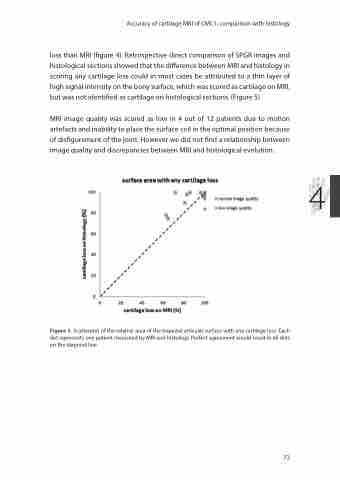
loss than MRI (figure 4). Retrospective direct comparison of SPGR images and histological sections showed that the difference between MRI and histology in scoring any cartilage loss could in most cases be attributed to a thin layer of high signal intensity on the bony surface, which was scored as cartilage on MRI, but was not identified as cartilage on histological sections. (Figure 5)
MRI image quality was scored as low in 4 out of 12 patients due to motion artefacts and inability to place the surface coil in the optimal position because of disfigurement of the joint. However we did not find a relationship between image quality and discrepancies between MRI and histological evolution.
4
Figure 3. Scatterplot of the relative area of the trapezial articular surface with any cartilage loss. Each dot represents one patient measured by MRI and histology. Perfect agreement would result in all dots on the diagonal line.
73
