
Page 98 - Magnesium-based supports for stem cell therapy of vascular disease - Mónica Echeverry Rendón
P. 98
CHAPTER 6
dexamethasone (Pharmacy UMCG, Netherlands), 1nM of insulin (Gibco, 41400-045, Waltham, Massachusetts, USA), 0.05mM 3-isobutyl-1-methylxanthine (IBMX) (Sigma-Aldrich I5879, St. Louis, Missouri, USA). Cells were stained with oil red solution. For osteoblastic differentiation, ASC were exposed to 0.1μM of dexamethasone (Pharmacy UMCG), 10 mM of β-glycerophosphate (Sigma, G9891, Sigma-Aldrich, St. Louis, Missouri, USA), 0.05mM ascorbic acid (Sigma, A4544, Sigma-Aldrich, St. Louis, Missouri, USA). Cells were stained with alizarin red. For adipogenic and osteogenic staining, cells were assessed with an inverted optical microscope Leica DMRXA microscope and Leica software. Finally, for smooth muscle differentiation, ASC were treated with 5 ng/ml of Recombinant Hu- man TGF-β1 (Peprotech,NJ, USA) and after two weeks cells were fixed and stained with antibodies against SM22☆ (1:500, Abcam, ab14106, Cambridge, U.K), phalloidin (1:500, A12379, Thermo Fisher Scientific, Waltham, Massa- chusetts, USA), and DAPI (1:5000, Sigma-Aldrich, D9542, St. Louis, Missouri, USA). As secondary antibody Alexa fluor 647 donkey anti-rabbit IgG H&L (Abcam, ab150075, Cambridge, U.K) Cells were with a TissueFAXs microscope (TissueGnostics GmbH, Vienna, Austria).
2.9 Co-culture of ASC and HUVEC
To assess the influence of materials’ leachables from surface-coated materials and c.p Mg, the angiovasculogenic capacity of HUVEC on ASC monolayers in wells of a 24 well plate was determined in two manners. Firstly, ASC were exposed two materials’ leachables for 24h. Next, the medium was removed and a single cell suspension of HUVEC was seeded at 10,000 per cm2. The formation of vascular networks was monitored for seven days. Secondly, HUVEC were seeded on top ASC monolayers at 10,000 per cm2 and incubated for seven days to allow vascular networks to form. Next, the medium was replaced with materials’ leachables and further incubated for 24h. At the end of the experiment, for both types of treatment, cells were washed twice and fixed with 2% paraformaldehyde in PBS for 30 min. Cells were permeabilized with 0.5% Triton X-100 (Sigma-Aldrich, St. Louis, Missouri, USA) for 15 min. Next, cells were incubated with monoclonal mouse anti-human CD31 (1:100, Dako, Clone JC70A, California, USA), for detection of endothelial cells and rabbit polyclonal SM22 (1:100, Abcam, ab14106, Cambridge, U.K) for 2 hours. Then cells were washed with PBS and incubated with secondary antibodies Alexa Fluor 488 Donkey anti-Mouse IgG (H+L) (1:300, Life technologies, A-21202, California, USA) and Alexa Fluor 647 Donkey anti-Rabbit IgG (H+L) (1:300, Life technologies, A31573, California, USA) for 1h. Samples were preserved in PBS and observed using a TissueFAXs microscope (TissueGnostics GmbH, Vienna, Austria).
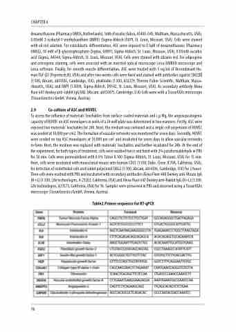
Table2.Primer sequence for RT-qPCR
96
