
Page 96 - Magnesium-based supports for stem cell therapy of vascular disease - Mónica Echeverry Rendón
P. 96
CHAPTER 6
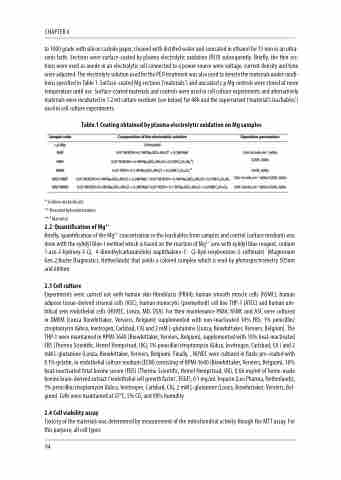
to 1000 grade with silicon carbide paper, cleaned with distilled water and sonicated in ethanol for 15 min in an ultra- sonic bath. Sections were surface-coated by plasma electrolytic oxidation (PEO) subsequently. Briefly, the thin sec- tions were used as anode in an electrolytic cell connected to a power source were voltage, current density and time were adjusted. The electrolyte solution used for the PEO treatment was also used to denote the materials under condi- tions specified in Table 1. Surface-coated Mg sections (‘materials’) and uncoated c.p Mg controls were stored at room temperature until use. Surface-coated materials and controls were used in cell culture experiments and alternatively materials were incubated in 1.2 ml culture medium (see below) for 48h and the supernatant (‘material’s leachables’) used in cell culture experiments.
Table.1 Coating obtained by plasma electrolytic oxidation on Mg samples
* Sodium metasilicate
** Hexamethylenetetramine
*** Mannitol
2.2 Quantification of Mg2+
Briefly, quantification of the Mg2+ concentration in the leachables from samples and control (culture medium) was done with the xylidyl blue-I method which is based on the reaction of Mg2+ ions with xylidyl blue reagent, sodium 1-azo-2-hydroxy-3-(2, 4-dimethylcarboxanilido)-naphthalene-1’- (2-hyd-roxybenzene-5-sulfonate) (Magnesium Gen.2,Roche Diagnostics, Netherlands) that yields a colored complex which is read by photospectrometry 505nm and 600nm.
2.3 Cell culture
Experiments were carried out with human skin fibroblasts (PK84), human smooth muscle cells (hSMC), human adipose tissue-derived stromal cells (ASC), human monocytic (premyeloid) cell line THP-1 (ATCC) and human um- bilical vein endothelial cells (HUVEC, Lonza, MD, USA). For their maintenance PK84, hSMC and ASC were cultured in DMEM (Lonza Biowhittaker, Verviers, Belgium) supplemented with non-inactivated 10% FBS, 1% penicillin/ streptomycin (Gibco, Invitrogen, Carlsbad, CA) and 2 mM L-glutamine (Lonza, Biowhittaker, Verviers, Belgium). The THP-1 were maintained in RPMI-1640 (Biowhittaker, Verviers, Belgium), supplemented with 10% heat-inactivated FBS (Thermo Scientific, Hemel Hempstead, UK), 1% penicillin/streptomycin (Gibco, Invitrogen, Carlsbad, CA ) and 2 mM L-glutamine (Lonza, Biowhittaker, Verviers, Belgium). Finally, , HUVEC were cultured in flasks pre-coated with 0.1% gelatin, in endothelial culture medium (ECM) consisting of RPMI-1640 (Biowhittaker, Verviers, Belgium), 10% heat-inactivated fetal bovine serum (FBS) (Thermo Scientific, Hemel Hempstead, UK), 0.06 mg/ml of home-made bovine brain-derived extract (‘endothelial cell growth factor’, ECGF), 0.1 mg/mL heparin (Leo Pharma, Netherlands), 1% penicillin/streptomycin (Gibco, Invitrogen, Carlsbad, CA), 2 mM L-glutamine (Lonza, Biowhittaker, Verviers, Bel- gium). Cells were maintained at 37°C, 5% CO2 and 98% humidity.
2.4 Cell viability assay
Toxicity of the materials was determined by measurement of the mitochondrial activity though the MTT assay. For this purpose, all cell types
94
